Title: Dynamic Thermal Imaging – A Valuable Measurement Method for Biomedical Applications
Abstract: Thermal imaging, or thermography, consists in measuring and imaging the thermal radiation emitted by every object above the absolute zero temperature. As this radiation is temperature-dependent, the infrared images recorded can be converted into temperature maps, or thermograms, allowing retrieving valuable information about the object under investigation. Thermal imaging has been known since the middle of the 20th century and recent technological achievements concerning the infrared imaging devices, together with the development of new procedures based on transient thermal emission measurements revolutionized the field. Nowadays, thermography is a method whose advantages are undisputed in engineering. It is routinely used for the non-destructive testing of materials, to investigate electronic components, or in the photovoltaic industry to detect defects in solar cells.
Despite an early interest, thermal imaging is currently rarely used for biomedical applications and even less in clinical settings. One reason is probably the initial disappointing results obtained solely with static measurement procedures, where the sample is investigated in its steady state, and using unhandy and performance-limited first-generation infrared cameras. In addition, the retrieval of quantitative data using dynamic thermal imaging procedures often requires complex mathematical modelling of the sample which can be demanding in biomedical applications due to the large variability intrinsic to life science field.
The goal of this lecture is to a) demonstrate the potential of dynamic thermal imaging for biomedical applications and b) give the reader the necessary background to successfully translate the technology to his/her specific biomedical applications.
In a first step, the basics of thermal radiation and thermal imaging device technology will be reviewed. Rather than giving an exhaustive description of the technology, we aim to familiarize the reader with key concepts that will allow selecting an optimal infrared camera depending on the specific application. In a subsequent part, we will present the foundation of thermodynamics needed to understand and be able to mathematically model heat transfer processes happening inside the sample under investigation and between the sample and its environment. Such thermal exchanges are responsible for the sample surface temperature. As next steps, we will present in detailed and compare the different procedures used in dynamic thermal imaging. Dynamic thermal imaging means that the sample surface thermal emission is monitored in its transient state and exhibit superior capabilities compared to passive thermal imaging. The thermal stimulation can be achieved with different modalities depending on the sample under investigation (LASER or flash lamps to investigate thin coatings, alternating magnetic fields to detect magnetic material, microwave to heat up water, or ultrasound to monitor cracks). Various procedures are possible: stepped- and pulsed-thermal imaging, pulsed-phase and lock-in thermal imaging. Each approach exhibiting specific characteristics in term of signal to noise ratio or measurement duration.
As an illustration, we will demonstrate how lock-in thermal imaging can be advantageously used to build extremely sensitive instruments to detect and characterize stimuli-responsive nanoparticles (both plasmonic and magnetic) in complex environments like cell cultures, tissue or food. In this example, we will present the research instrument in detail with the choice of the various components, the digital lock-in demodulation implemented, the mathematical modelling of the sample required to extract quantitative information as well as the resulting setup performances. The goal being to allow the reader to translate the dynamic thermal imaging measurement principles to its own biomedical application.
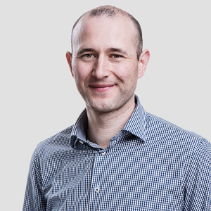
Bio: Mathias Bonmarin studied at the graduate school of science and engineering “POLYTECH Marseille” part of Aix-Marseille University (France) and obtained his master’s degree in biomedical engineering in 2002. He then moved to Grenoble Institute of Technology to gain an additional master’s in optics, optoelectronics, and microwave. After a year working as a scientific assistant at the Fresnel Institute in Marseille, he decided to broaden his perspective and studied economics at the University of Louvain-La-Neuve in Belgium where he graduated “summa cum laude.” Mathias moved to Switzerland in 2006 to do his Ph.D. in physical chemistry at the University of Zurich. He joined the School of Engineering of the Zurich University of Applied Science (ZHAW) as a research associate in 2010 before being appointed senior lecturer at the Institute of Computational Physics in 2014. Between July 2018 and July 2019, Mathias was a Fulbright visiting research scholar at the College of Engineering and Applied Sciences of the University of Cincinnati in the US.
Since July 2019 he is a Professor of optoelectronics and leads the Sensors and Measurement Systems group at ZHAW. His research interests focus on the development of innovative sensors and instruments for medicine and biology. Mathias is IEEE senior member (IMS and EMBS) and received the 2019 Best Application in I&M Award.
Please click this URL to start or join. https://iastate.zoom.us/j/96810972944?pwd=SVVLWlY2cVdZYXhxWWg4ZHF1cVdSZz09
Or, go to https://iastate.zoom.us/join and enter meeting ID: 968 1097 2944 and password: 334840
Join from dial-in phone line:
Dial: +1 309 205 3325 or +1 312 626 6799
Meeting ID: 968 1097 2944
Participant ID: Shown after joining the meeting
International numbers available: https://iastate.zoom.us/u/aqUgrVklM